Clinical service and research

The laboratory for movement examinations is part of the Neuroorthopaedics of the University Children’s Hospital of Basel. The task of the laboratory is a symbiosis between clinical service and research. The working group investigates the effects of therapies on walking, especially orthotic fittings or surgical interventions. Clinical research questions usually originate in the clinic, the consultation hour, physiotherapy or the operating theatre. The research results and the resulting new findings flow back into clinical examination methods and treatments.

In the future, valuable information on pathological walking from musculoskeletal simulations and the finite element method.

The group is also working on the further development of its own kinematic foot model, which combines data from pedobarography, ground reaction force measurement and foot X-ray images.

Equipment Used

Vantage is Vicon’s flagship range of cameras. The sensors have resolutions of 5, 8 and 16 megapixels, with sample rates up to 2000Hz – this allows you to capture fast movements with very high accuracy. The cameras also have built-in temperature and bump sensors, as well as a clear display, to warn you if cameras have moved physically or due to thermal expansion. High-powered LEDs and sunlight filters mean that the Vantage is also the best choice for outdoor use and large volumes.

The Cometa Pico (EMG) sensors are small, light-weight and have on-board storage to allow measurements in the field. Easy to attach and easy to charge, the long battery life, the high signal-to-noise ratio and the wireless range are other features that make the aktos the best EMG system on the market today.

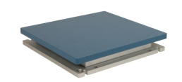
The Optima is the flagship among force plates. The patented calibration technology guarantees the highest possible accuracy across the entire surface of the plate – ideal for gait analysis, biomechanical research and other applications where the highest quality data is essential.

Special project UKBB

We are particularly proud of our scientific project to visualise specific leg muscles using 3D ultrasound to study muscle morphology. To do this, we synchronised our Vicon system with an ultrasound device. The project was conducted in collaboration with partners at VU (Vrije Universiteit) University Medical Center Amsterdam (NL) and Vrije Universiteit Amsterdam (NL) and the joint publication (PLoS ONE 11(11): e0166401. doi:10.1371/journal.pone.0166401) won the 2017 Science Award of the Association for Paediatric Orthopaedics (VKO), a section of the German Society for Orthopaedics and Orthopaedic Surgery (DGOOC).

Are you interested in a similar solution?

Ask here!